Up Close and Unmatched: New Microscope a First-of-Its-Kind in the Region
A recent major investment in Syracuse University research infrastructure has resulted in the installation of a Zeiss Sigma 360 field emission scanning electron microscope in the University’s Materials Research Core (MRC) facility. The instrument has introduced dramatic new imaging capabilities to researchers at the University and at partner institutions in the region.
The new instrument demonstrates the University’s commitment to supporting and enabling cutting-edge research in important fields like biomedical engineering, materials science and quantum computing, says Jeremy Steinbacher, director of research operations in the Office of Research.
The Zeiss will serve researchers across disciplines and career stages, from advanced undergraduates and graduate students to postdoctoral scholars and faculty. The Zeiss also supports the campus Quantum Information Science research group and Central New York’s rapidly expanding semiconductor and quantum technology ecosystem. The instrument was funded by a $335,000 investment by the Office of Research, the BioInspired Institute and individual faculty contributors.

On Campus and Beyond
The microscope is part of the Office of Research’s efforts to build shared, core facilities available to users across the University and the greater Syracuse region, says Duncan Brown, vice president for research. “Strong core facilities are a force multiplier for our outstanding faculty and student researchers, providing access to state-of-the-art scientific instruments without the burden of having to purchase and maintain them individually.”

“For researchers who once drove an hour to use a scanning electron microscope, that capability is now right here, benefiting researchers on our campus, in our community and throughout the region,” Steinbacher says. It also serves as a recruiting tool because it demonstrates to prospective graduate students, postdoctoral scholars and faculty that state-of-the-art instrumentation is readily accessible at Syracuse, he says.
A Billionth of a Meter
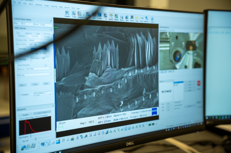
Its resolution of 1.6 nanometers means the Zeiss can zoom down to the nanoscale, revealing details as small as a billionth of a meter, sharp enough to capture images of computer chip components, nanoparticles, bacteria and living cells, Steinbacher says.
It captures the shape and texture of an object’s surface in detailed, three-dimensional images versus thin cross-sections of materials. Because its electron beam works at lower energy levels, the microscope also offers highly detailed viewing of soft or non-metallic materials that typically are difficult or impossible to examine with older equipment, Steinbacher says.
Conventional electron microscopes require samples to be stripped of all moisture and placed under high vacuum, but some materials fall apart or change when dried out. Zeiss permits variable pressure imaging, so air pressure inside the imaging chamber can be adjusted to view samples that aren’t bone-dry. That lets researchers examine hydrogels, drug-delivery particles and biological samples in a more natural state. That capacity did not previously exist at Syracuse University or other area institutions, according to Steinbacher.
Who Will Use It
Biomedical and chemical engineering researchers can use the microscope to examine polymer film morphology. Environmental scientists can image rocks and fossils. Others will use it for battery technology research and catalyst design. The Quantum Information Science group and scientists in electrical engineering, computer science and physics can conduct device characterization—testing device effectiveness and checking for flaws.
Eric Finkelstein, technical director of the Materials Research Core, says the Zeiss enables exciting new levels of research. “It lets researchers image the surface appearance of synthetic materials, such as polymers or other engineered materials, and biological samples, such as cells, tissues and organisms, at higher resolution and better definition compared to existing instruments in the area.”
The instrument “is a critical addition to Syracuse’s growing suite of fabrication and characterization tools for next-generation quantum technologies,” says Ethan Arnault, assistant professor of electrical engineering and computer science in the College of Engineering and Computer Science. “We’ll use it to image our superconducting devices at the nanometer scale, hunting down the surface defects and contaminants that limit their performance.”
Ivan Pechenezhskiy, assistant professor of physics in the College of Arts and Sciences, says the Zeiss will assist in prescreening superconducting qubit devices—the tiny, ultra-cold circuits that are the building blocks of quantum computers—from device batches fabricated elsewhere. “That will help us focus on the most promising devices and let students make the connection between the abstract shapes they draw on computer screens and the actual footprints of the tiny electrical circuits their designs imprint on the chips.”
For more information about access and use of University instruments and facilities, visit the Core Facilities webpage.
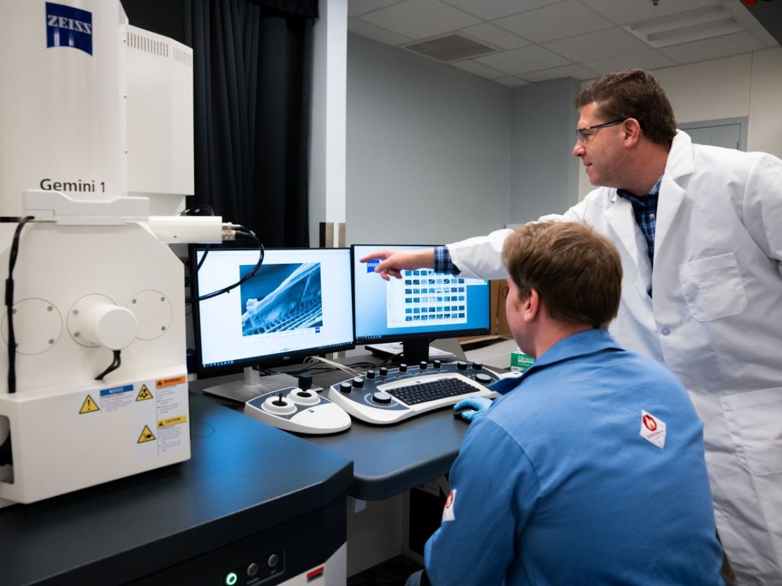
The Zeiss serves researchers across disciplines and career stages, from advanced undergraduates and graduate students to postdoctoral scholars and faculty. The instrument can zoom down to the nanoscale, revealing details as small as a billionth of a meter. (Photo by Amy Manley)
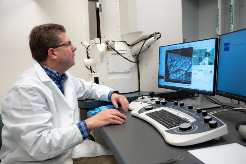
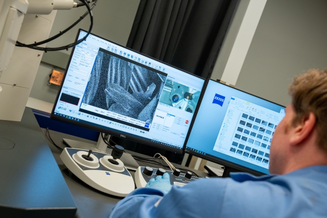
The new microscope captures the shape and texture of an object’s surface in detailed, three-dimensional images versus thin cross-sections of materials. (Photo by Amy Manley)
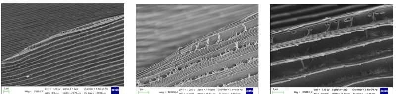
These images show the structure of a butterfly wing at progressively higher magnifications using the new Zeiss instrument. At left is the view at 2500 X (times) magnification and in the middle and at right, at 10,000 X. The repeating lines and patterns form the physical structure responsible for butterfly wing coloration. (Photo compiled by Eric Finkelstein)
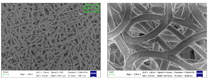
Close-ups of the polymer substance polycaprolactone, which is used in biomedical applications such as drug delivery and tissue engineering after electrospinning to produce fibers. The photo at left is at 300 X magnification. The photo at right, taken by the microscope’s in-lens detector, shows it at 2500 X magnification. The images illustrate the random orientation and uniform size of fibers that measure about two microns in diameter, as well as the surface topography of individual fibers. (Photo compiled by Eric Finkelstein)
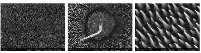
The Zeiss closeups show the skin of a gecko depicting the animal’s setae, bristle-like structures that enable its extraordinary adhesion to surfaces, water repellency and self-cleaning capabilities. At left the setae are shown at 2000 X (times) magnification and in the center photo, at 23,000 X. The image at right shows a highly magnified papillae (a hair-like structure) that contributes to the gecko skin’s antimicrobial and water repellent properties. (Photo compiled by Eric Finkelstein)